
The idea of being asked to go for a Mammogram Screening can be quite terrifying, especially because they are associated with cancer. They are not always to be feared though as they are there to keep tabs on your breasts and check for any lumps, bumps or abnormalities. If you were to get an early detection through a mammogram screening then it would mean treatment would be more effective. A mammogram screening can pick up abnormalities before the feeling of lumps and bumps become apparent to the human touch.
 What is a Mammogram Screening
A mammogram is an x-ray image of the breast that shows changes in the tissue, new lumps, or tiny clusters of calcium that can be the first signs of cancer that cannot be felt or seen on MRI, Ultrasound or breast self-exam. Mammography is the most effective method for detecting cancer in the early stages, and are often the first line of early detection. Additional screening methods may be needed to find out if abnormal cells are present. Routine screening mammograms can find the presence of cancerous cells before it grows or spreads which can greatly increase survivability. It is so important that you do attend your appointment when being asked to go for a mammogram screening, it could potentially be saving your life, to just dismiss it or not see the importance of it could be detrimental to you.
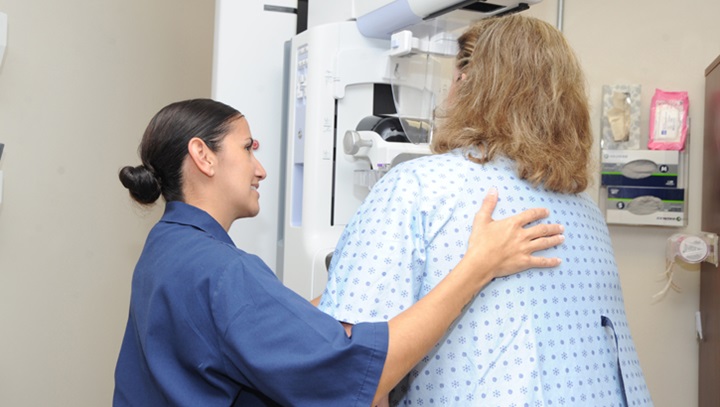
The Mammogram Screening Procedure
When you arrive to have your mammogram screening you will be asked to undress from above the waist and you will be given a gown to wear. The nurse will bring you to the mammography room, where our mammography technician will help position your breast between two flat panels that are used to gently compress the breast to flatten and spread out the breast tissue. Whilst your breast is being compressed you are likely to experience a bit of discomfort but just remember that the compression part of the mammogram screening is very important. Compressing the breast thins and evens the breast tissue and is necessary to obtain clear x-rays images. The mammography technician will then leave the room or go behind a screen and begin taking the x-rays. This process of positioning and imaging is repeated for additional views of the same breast or the other breast. The entire procedure should take between 10 to 20 minutes for conventional mammography and a shorter 5 to 10 minutes for digital mammography.
Receiving Abnormal Screening Results
If your results return with abnormalities then additional imaging studies may help clarify the abnormality and determine whether biopsy or follow-up imaging in 6 months is required. If the abnormalities are likely benign, repeat mammogram or ultrasound may be recommended in 6 months to document stability. Generally, if an abnormality does not change in a year, it is considered benign. At this stage of early diagnosis, should the abnormality prove to be malignant, there is an excellent chance of cure.